VASCULAR MALFORMATIONS – HEMANGIOMA
Introduction
Vascular malformations usually develop during embryonic life. In most cases, these malformations gradually enlarge after birth and may cause cosmetic or functional problems, such as pain or swelling. Vascular malformations appear as reddish lesions caused by the abnormal development of blood vessels, including capillaries, veins, arteries, or lymphatic vessels. They may occur anywhere in the body.
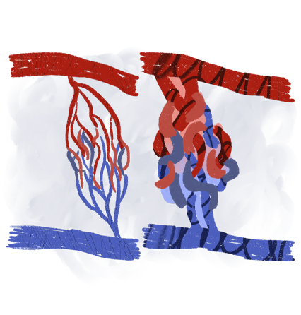
Hemangiomas, on the other hand, are benign vascular tumors that typically appear after birth. They usually present as red nodules or plaques on the skin. Their size can vary, and diagnosis is usually based primarily on the clinical appearance of the lesion. Depending on their size and location, hemangiomas can be treated conservatively, surgically, or with laser therapy.
The timely early diagnosis of the disease
is very important
for the good outcome of the condition in both stages of the disease.
Types of malformations
Vascular malformations are classified into four main categories:
Capillary malformations involve the smallest blood vessels in the body, known as capillaries, and are most commonly found on the face. They rarely cause medical symptoms, and treatment is usually indicated mainly for cosmetic reasons.
Venous malformations develop within abnormally formed veins and may appear in various parts of the body. Symptoms depend on the location and extent of the malformation. A particularly extensive form of venous malformation is Klippel–Trenaunay syndrome.
Lymphatic malformations usually develop in the lower limbs and can cause significant swelling (edema) and cystic abnormalities.

Diagnosis of Vascular Malformations
Diagnosis is based on clinical examination combined with imaging techniques, including Duplex ultrasound (Triplex), magnetic resonance angiography (MRA), and computed tomography angiography (CTA). These imaging methods allow detailed assessment of the extent and type of vascular malformation.
Ποιες είναι οι επεμβατικές μέθοδοι χειρουργικής θεραπείας;
Treatment of Vascular Malformations
Treatment of vascular malformations is complex and requires careful planning. Patients should be aware that, depending on the size and complexity of the malformation, multiple treatment sessions may be required to achieve optimal results.
Capillary malformations, particularly those on the face, are usually treated with laser therapy.
Arteriovenous malformations are initially treated using minimally invasive endovascular techniques, usually through embolization of the arterial branches supplying the malformation. If necessary, embolization of the venous branches may follow at a later stage.
Veno-venous malformations are treated through direct embolization using specialized sclerosing agents, such as ethanol and polidocanol (Aethoxysklerol). Because of the risk of recurrence or the need for large quantities of sclerosing agents, an advanced technique called electroporation with simultaneous administration of the drug bleomycin may be used, particularly for extensive malformations of the body or face. In this technique, electrical impulses temporarily open the cell membranes, allowing the medication to enter the cells more effectively. This increases the drug’s effectiveness by approximately 100 times, leading to the destruction of abnormal vascular cells and gradual regression of the malformation. The same technique has also shown excellent results in lymphatic malformations.