Aortic Arch Aneurysm
Introduction
An aortic arch aneurysm is a serious medical condition due to the risk of rupture. The aorta is the largest artery in the human body and carries blood from the heart to the rest of the body. The aortic arch is the segment of the aorta located between the ascending aorta and the descending aorta. Its main function is to supply blood to the arteries that feed the brain and the upper limbs.
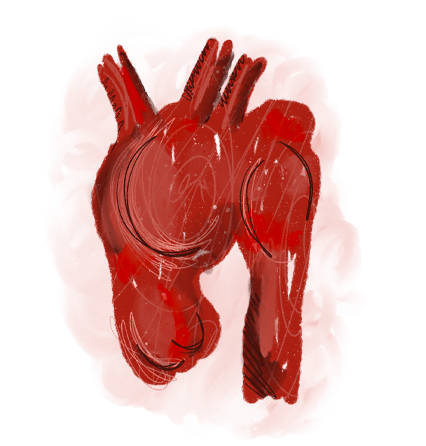
When an aneurysm develops in the aortic arch, early diagnosis is critically important, as rupture can have life-threatening consequences. Unfortunately, most aortic arch aneurysms are asymptomatic and are often discovered incidentally during imaging tests, such as a chest X-ray or ultrasound performed for other medical reasons.
Ποιος την αντιμετωπίζει: Ο Θεοδόσιος Μπίσδας, MD, PhD, FACS, Αν. Καθηγητής Αγγειοχειρουργικής και Διευθυντής της Γ’ Αγγειοχειρουργικής Κλινικής του Ιατρικού Κέντρου Αθηνών, εξειδικεύεται στην ενδαγγειακή αποκατάσταση του ανευρύσματος αορτικού τόξου (TEVAR). Πραγματοποίησε την πρώτη στην Ελλάδα ενδαγγειακή αποκατάσταση ανευρύσματος αορτικού τόξου με το ενδομόσχευμα Nexus Duo, χωρίς στερνοτομή, στο υβριδικό χειρουργείο του Ιατρικού Κέντρου Αθηνών.


The timely early diagnosis of the disease
is very important
for the good outcome of the condition in both stages of the disease.
Ανεύρισμα αορτικού τόξου: Χειρουργικές μέθοδοι
Understanding Aortic Arch Aneurysms
The aortic arch is the curved portion of the aorta that begins at the aortic valve and bends backward before continuing as the thoracic aorta, which runs through the chest in front of the spine.
The most common causes of aortic arch aneurysms include aortic dissection (Type A), smoking, arterial hypertension, and genetic connective tissue disorders such as Marfan syndrome or Ehlers–Danlos syndrome.
If left untreated, an aneurysm of the aortic arch—whether caused by aortic dissection or progressive dilation of the vessel wall—may lead to rupture, which is the most serious complication and can result in death.
Treatment Options for Aortic Arch Aneurysm
Traditionally, aortic arch aneurysms have been treated with open surgical repair, usually performed through sternotomy by specialized cardiac surgery teams. In recent years, however, minimally invasive endovascular techniques using stent grafts have been developed with excellent clinical outcomes, particularly in patients who have previously undergone sternotomy. Endovascular repair may be indicated in selected cases (when anatomy allows), including:
- Dissecting or non-dissecting aneurysms of the aortic arch in patients who have already undergone replacement of the ascending aorta
- Saccular aneurysms of the aortic arch
- Aneurysms of the aortic arch or thoracic aorta with a normal ascending aorta

The role of the vascular surgeon
The minimally invasive treatment of aortic arch aneurysms is technically demanding and is offered by only a limited number of specialized centers worldwide. Our vascular surgery department at Athens Medical Center is among these centers. With extensive experience in the treatment of complex vascular diseases, vascular surgeon Dr. Theodosios Bisdassafely manages each case using advanced endovascular techniques. The most important factor in selecting the optimal treatment is the presence of a multidisciplinary Aortic Team. Within this team, cardiothoracic surgeons and vascular surgeons jointly evaluate each case, discuss the diagnosis and treatment strategy, and determine the safest and most effective intervention for the patient..
Frequently Asked Questions (FAQs)
What is the aortic arch?
The aortic arch is the section of the aorta that begins after the ascending aorta and curves backward and downward. From it arise the large vessels that supply blood to the brain, neck and upper limbs: the innominate artery, the left carotid artery and the left subclavian artery. It constitutes a central key point of circulation.
What does "aortic arch prominence" or "aortic knob prominence" mean?
The term "prominence" describes the impression that the aortic arch gives on a chest X-ray when it projects more than normal. The aortic knob is the shadow of the aortic arch on the left border of the heart. The prominence may be a normal finding in elderly individuals or may be associated with pathological conditions, such as hypertension or aneurysm.
What is calcification or calcification of the aortic arch?
Calcification of the aortic arch is the deposition of calcium in the wall of the aorta. It occurs frequently in elderly individuals or in patients with atherosclerosis, hypertension and diabetes mellitus. Although it may be an incidental finding on imaging, it indicates chronic damage to the vascular wall and increased cardiovascular risk.
What are the normal dimensions of the ascending aorta and the aortic arch?
The ascending aorta normally has a diameter of approximately 2.5–3.5 centimeters in adults, with small variations depending on sex and body type. The aortic arch has a slightly smaller diameter, around 2.5–3 centimeters. When the dimensions exceed 4 centimeters it is considered a disorder, while above 5 centimeters there is an indication for monitoring or treatment.Sonnet 4.6
How is the aortic arch visualized on an X-ray?
On a plain chest X-ray, the aortic arch forms the well-known "aortic knob", that is a curved shadow on the left outline of the mediastinum. Its prominence may become more apparent in individuals with hypertension, age-related changes or an aneurysm. For accurate measurement of dimensions, examinations such as CT scan or MRI are required.Sonnet 4.6
How is the aortic arch treated?
The treatment of the aortic arch depends on whether there is a pathology, such as an aneurysm or dissection. Traditionally, treatment was performed with open surgery, which remains the most radical method. However, in recent years new endovascular techniques have been developed (TEVAR – Thoracic Endovascular Aortic Repair), where endoprostheses are placed with a catheter via the femoral artery. These methods are less invasive, reduce recovery time and offer solutions to high-risk patients. The choice of method depends on the size, location, general health of the patient and the experience of the medical team.
Ποιος αγγειοχειρουργός στην Αθήνα αντιμετωπίζει το ανεύρυσμα αορτικού τόξου ενδαγγειακά;
Ο Θεοδόσιος Μπίσδας, Αν. Καθηγητής Αγγειοχειρουργικής και Διευθυντής της Γ’ Αγγειοχειρουργικής Κλινικής του Ιατρικού Κέντρου Αθηνών, εξειδικεύεται στην ενδαγγειακή αντιμετώπιση των παθήσεων του αορτικού τόξου. Πραγματοποίησε την πρώτη στην Ελλάδα ενδαγγειακή αποκατάσταση ανευρύσματος αορτικού τόξου με το ενδομόσχευμα Nexus Duo, χωρίς στερνοτομή, στο υβριδικό χειρουργείο του Ιατρικού Κέντρου Αθηνών.
Τι είναι η ενδαγγειακή αποκατάσταση (TEVAR) του αορτικού τόξου;
Η TEVAR (Thoracic Endovascular Aortic Repair) είναι η ελάχιστα επεμβατική τοποθέτηση ενδομοσχεύματος στη θωρακική αορτή μέσα από μικρές τομές στη μηριαία αρτηρία, χωρίς ανοιχτό χειρουργείο. Στο αορτικό τόξο εφαρμόζεται με ειδικά διακλαδισμένα ενδομοσχεύματα, όπως το Nexus, που διατηρούν την αιμάτωση των αγγείων του εγκεφάλου. Προσφέρει ταχύτερη ανάρρωση και σημαντικά μικρότερη επιβάρυνση σε σχέση με τη στερνοτομή.
Τι είναι το ενδομόσχευμα Nexus Duo για το αορτικό τόξο;
Το Nexus Duo είναι ενδομόσχευμα νέας γενιάς, ειδικά σχεδιασμένο για την ενδαγγειακή αποκατάσταση του αορτικού τόξου μετά από ανεύρυσμα ή χρόνιο διαχωρισμό. Τοποθετείται χωρίς στερνοτομή και διατηρεί τη ροή προς τους κλάδους του αορτικού τόξου. Η πρώτη εμφύτευσή του στην Ελλάδα πραγματοποιήθηκε από τον Θεοδόσιο Μπίσδα στο υβριδικό χειρουργείο του Ιατρικού Κέντρου Αθηνών. Διαβάστε περισσότερα στη σελίδα για το ενδομόσχευμα Nexus Duo.
Πότε χρειάζεται χειρουργείο ένα ανεύρυσμα αορτής;
Κατά κανόνα, επέμβαση συνιστάται όταν η διάμετρος του ανευρύσματος υπερβαίνει τα 5-5,5 εκατοστά, όταν αυξάνεται γρήγορα (περισσότερο από 0,5 εκατοστά το εξάμηνο) ή όταν προκαλεί συμπτώματα. Η απόφαση εξατομικεύεται από τον αγγειοχειρουργό με βάση τη θέση και τη μορφολογία του ανευρύσματος και τη γενική κατάσταση του ασθενούς.