Deep Vein Thrombosis (DVT)
Introduction
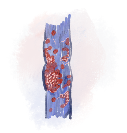
Venous thrombosis refers to the formation of blood clots within the venous system. Depending on where the clot forms—either in the superficial veins or the deep veins—the condition is classified as superficial venous thrombosis or deep vein thrombosis (DVT). Blood clots obstruct the normal flow of blood through the veins and, in some cases, may also affect arterial circulation.

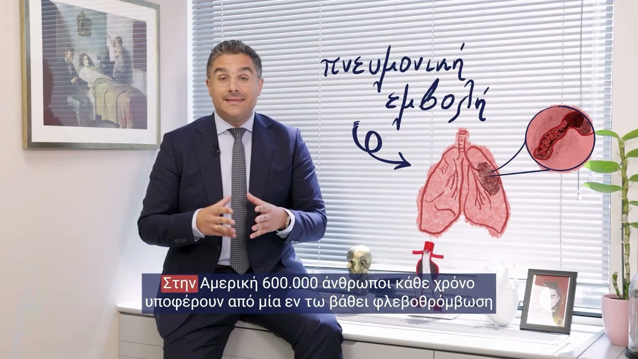
Although approximately 50% of patients with venous thrombosis are asymptomatic, the most common symptoms include pain, swelling, skin tenderness at the site of the clot, redness, and a burning sensation. If venous thrombosis is not diagnosed and treated early, the clot may migrate toward the heart and cause a pulmonary embolism, a potentially life-threatening complication.
Ποιος την αντιμετωπίζει: Ο Θεοδόσιος Μπίσδας, Αν. Καθηγητής Αγγειοχειρουργικής και Διευθυντής της Γ’ Αγγειοχειρουργικής Κλινικής του Ιατρικού Κέντρου Αθηνών, αντιμετωπίζει την εν τω βάθει φλεβική θρόμβωση (DVT) τόσο με φαρμακευτική αγωγή όσο και με σύγχρονες ενδαγγειακές τεχνικές θρομβοαναρρόφησης στο υβριδικό χειρουργείο του Ιατρικού Κέντρου Αθηνών, όταν απαιτείται άμεση αφαίρεση του θρόμβου.


The timely early diagnosis of the disease
is very important
for the good outcome of the condition in both stages of the disease.
Understanding Venous Thrombosiss
The venous thrombosis can be caused if at least one of the following factors is present in the veins and blood of the patient:
(1) alteration of the composition of the blood (e.g. thrombophilia, cancer)
(2) damage to the inner wall of the veins (e.g. stent, catheter injury, venous insufficiency, stenosis
(3) obstruction of flow with blood stasis (e.g. Chronic venous insufficiency, vein compression, thoracic inlet/outlet syndrome, May-Thurner syndrome).
Depending on the location of the clot there are two types of venous thrombosis: superficial thrombophlebitis (superficial thrombophlebitis) and the Compared with deep vein thrombosis (DVT),. The superficial thrombophlebitis is distinguished by intense local pain along the vein, redness and a feeling of hardness. The Compared with deep vein thrombosis (DVT), has more intense symptoms with severe pain especially upon compression, swelling and redness of the lower limb, while in rarer cases the entire limb may be ischemic with unbearable pain.
The venous thrombosis is associated with several risk factors among which venous insufficiency, obesity, cancer, the combination of hormonal therapies with smoking, prolonged immobility, soft tissue injuries, thrombophilia etc. are included. A special case is May Thurner syndrome where at the level of the navel the left iliac vein is compressed by the right iliac artery. At this point both the aorta and the inferior vena cava bifurcate and the vessels of the bifurcation intersect. Finally, special mention must be made of thoracic outlet/inlet syndrome where the subclavian vein is compressed by the first rib and the muscles surrounding it. Thrombosis of the vein is commonly encountered in patients who do body building due to muscle hyperplasia from weights.
Ποιες είναι οι επεμβατικές μέθοδοι χειρουργικής θεραπείας;
Diagnosis and Treatment
The diagnosis of venous thrombosis is initially clinical in combination with the measurement in the blood of D-dimers. In the case where D-dimers are negative the patient definitely does not have thrombosis. In the opposite case, i.e. with elevated D-dimer levels, there is an increased probability of the presence of venous thrombosis. This is followed by the performance of a venous triplex where the presence or absence of a clot in the limbs or abdomen is confirmed. If there is clinical suspicion of pulmonary embolism, a CT angiography of the pulmonary arteries should be performed with or without a cardiac ultrasound.
The treatment of acute venous thrombosis is as a rule conservative with the administration of anticoagulant medications. If the thrombosis involves superficial veins the duration of treatment lasts approximately 45 days while in cases of deep venous thrombosis the duration of treatment is decided based on the underlying cause with a minimum duration of 3 to 6 months.
In the case where the thrombosis is located in the veins of the abdomen and is acute (<14 days) it can now be treated with special minimally invasive clot removal techniques. Timely removal offers the advantage of immediate improvement of symptoms but also the avoidance of the so-called post-thrombotic syndrome (permanent swelling in the limbs, skin color change at the level of the ankles, appearance of venous ulcer). This technique of thromboaspiration is also recommended in cases of massive pulmonary embolisms with cardiorespiratory insufficiency for the patient and risk of death.
In cases of older thromboses in the large veins of the abdomen with the appearance of symptoms of post-thrombotic syndrome (see above) minimally invasive opening of the vessels is recommended using special venous endografts (stents). Successful opening allows the decompression of the limb and the immediate improvement of symptoms.
With the Hybrid Clinic the opportunity is given for safe and bloodless treatment of venous thrombosis, without the presence of complications.

The role of the vascular surgeon
The key to successful treatment of venous thrombosis is early diagnosis and prompt management.Subsequently the Vascular Surgeon sets a plan of diagnostic tests, monitoring with triplex and anticoagulant therapy. Particularly in the case of central thromboses in the abdomen it is important for the vascular surgeon to intervene as soon as possible and aspirate the clot. The same applies to pulmonary embolism. In the special case of subclavian vein thrombosis in the context of thoracic outlet/inlet syndrome the opening of the vein and the immediate surgical removal of the first rib is recommended. Contact with our clinic for further information.
Frequently Asked Questions (FAQs)
What is venous thrombosis;
The venous thrombosis is the condition in which a blood clot (coagulated blood) forms inside a vein, obstructing the normal flow of blood.
Why is venous thrombosis dangerous;
When a clot blocks blood flow in a deep vein of the leg or arm, complications may range from mild symptoms such as swelling and pain to serious conditions like pulmonary embolism.
What types of venous thrombosis exist?
There are two main types of venous thrombosis: the deep venous thrombosis (DVT), which involves the deeper veins, and the superficial thrombophlebitis, which is located in the superficial veins just below the skin. Each type requires a different diagnostic approach and treatment with DVT being the most serious and dangerous form.
How can I tell if I have a blood clot in my leg?
The thrombosis in the leg (DVT) often manifests with unilateral swelling, pain or tenderness (usually in the calf or thigh), accompanied by warm, red or darkened skin and swollen or painful veins. In some cases the symptoms may be mild or completely absent, a fact that makes the Doppler ultrasound essential to confirm or rule out the diagnosis.
What is the role of D-dimers in diagnosis?
D-dimers are protein fragments that appear in the blood when a clot is being broken down. Elevated levels suggest that clot formation and breakdown are occurring in the body. However, they do not always indicate thrombosis; therefore, ultrasound imaging is required for confirmation.
What is superficial thrombophlebitis?
Superficial thrombophlebitis is inflammation of superficial veins accompanied by clot formation, causing pain, redness, warmth, and a palpable hardened vein beneath the skin. It is usually a benign and self-limiting condition, although anticoagulation or surgery may be required if the clot extends toward deep veins.
What complications can venous thrombosis cause?
The venous thrombosis can cause serious complications, such as pulmonary embolism, when the clot detaches and reaches the lungs, causing a blockage that can be life-threatening. Furthermore, it can lead to chronic venous insufficiency or post-thrombotic syndrom which is characterized by edema (swelling) of the leg, hyperpigmentation (brown or black color) of the ankles and appearance of ulcer if not treated in time.
How is venous thrombosis treated?
The superficial thrombophlebitis is treated conservatively with the administration of anticoagulant injection for a short period of time (usually 45 days). In the case where the clot approaches the entry point of the superficial vein of the leg into the deep venous system surgical ligation of the vein is recommended for the prevention of DVT.
The treatment to deep venous thrombosis can be performed either with the administration of anticoagulants (usually for 6 months) and frequent follow-up checks or with a minimally invasive method and aspiration of the clot. The choice of treatment depends on the extent of the clot, its location, the severity of the symptoms, the underlying condition and whether in the case of pulmonary embolism the cardiac and respiratory function of the patient is affected. The endovascular treatment includes the mechanical thrombectomy (removal of the clot) and the possible use of a stent if deemed necessary.
Σε ποιον αγγειοχειρουργό να απευθυνθώ για φλεβοθρόμβωση (εν τω βάθει φλεβική θρόμβωση) στην Αθήνα;
Ο Θεοδόσιος Μπίσδας, Αν. Καθηγητής Αγγειοχειρουργικής και Διευθυντής της Γ’ Αγγειοχειρουργικής Κλινικής του Ιατρικού Κέντρου Αθηνών, αντιμετωπίζει τη φλεβοθρόμβωση τόσο συντηρητικά όσο και με σύγχρονες ενδαγγειακές τεχνικές θρομβοαναρρόφησης στο υβριδικό χειρουργείο του Ιατρικού Κέντρου Αθηνών, όταν απαιτείται άμεση αφαίρεση του θρόμβου.
Πώς αντιμετωπίζεται σήμερα η εν τω βάθει φλεβική θρόμβωση (DVT);
Η βάση της θεραπείας είναι η αντιπηκτική αγωγή. Σε εκτεταμένη λαγονομηριαία θρόμβωση, σε έντονα συμπτώματα ή σε απειλή του σκέλους, εφαρμόζονται ενδαγγειακές τεχνικές μηχανικής θρομβεκτομής (θρομβοαναρρόφηση), που αφαιρούν τον θρόμβο μέσω καθετήρα και μειώνουν τον κίνδυνο μεταθρομβωτικού συνδρόμου.
Τι είναι η θρομβοαναρρόφηση (μηχανική θρομβεκτομή) στη φλεβοθρόμβωση;
Είναι ελάχιστα επεμβατική τεχνική κατά την οποία ειδικός καθετήρας εισάγεται στη φλέβα και αναρροφά τον θρόμβο, αποκαθιστώντας άμεσα τη ροή του αίματος. Πραγματοποιείται στο υβριδικό χειρουργείο με απεικονιστική καθοδήγηση και συχνά επιτρέπει την αποφυγή της παρατεταμένης θρομβόλυσης και των επιπλοκών της.