Category: Case Studies
Case Study 3: Cervical Hemangioma
Extensive cervical hemangioma in a young woman – successful treatment with BEST (Bleomycin Electrosclerotherapy).
Patient profile
Female, 23 years old, student.
Symptoms and duration
The patient had a visible hemangioma on her neck since childhood. In recent years, it showed progressive growth, resulting in visible deformity and frequent episodes of pain and inflammation. The aesthetic impact was significant, causing social embarrassment and low self-esteem.
Diagnosis
The diagnosis was made through clinical examination and contrast-enhanced magnetic resonance imaging (MRI), which revealed an extensive soft tissue hemangioma in the cervical region.
Therapeutic approach
Due to the size and location of the lesion, surgical removal was not feasible without significant consequences. The method of choice was BEST (Bleomycin Electrosclerotherapy), in which our team has extensive experience.
Treatment description
Under general anesthesia, a small dose of bleomycin was injected into the hemangioma. Subsequently, a controlled electric field was applied using specialized electrodes, which enhanced the drug’s penetration and caused selective destruction of the vascular tissue. The session lasted 90 minutes, and no complications occurred.
The patient underwent a total of 2 sessions over a period of 9 months.
Course and recovery
After each session, there was mild swelling that subsided within 48–72 hours. At the 6-month follow-up, it was observed that a significant shrinkage of the hemangioma and improvement in the aesthetic appearance. After completing the treatments, the lesion almost disappeared, with an excellent functional and aesthetic outcome.
What made it unique?
The case involved a young woman with an extensive lesion in a sensitive area, where surgical intervention would have carried significant risks. The BEST method offered a safe, effective, and minimally invasive solution, leading to an excellent outcome with minimal complications a safe and effective treatment with an impressive aesthetic outcome.
Message to the public:
Hemangiomas are not just an aesthetic issue – they can cause pain and functional problems. The BEST method, when applied by a specialized team, is an innovative and safe solution that can significantly improve patients' lives.
Case Study 2: Aortic Arch Aneurysm
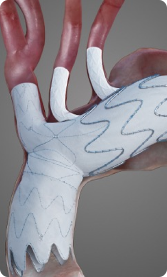
Endovascular repair of an aortic arch aneurysm using the Nexus stent graft – first application in Greece.
Patient profile
Male, 72 years old, with a significant cardiovascular history.
Symptoms and duration
The patient had a known Type A dissection which had been treated 5 years earlier with replacement of the ascending aorta. During long-term follow-up, a gradual dilation of the aortic arch was noted, reaching a diameter of over 6 cm. In the last six months, he reported chest pain and easy fatigue.
Diagnosis
The CT angiography revealed an aneurysm of the entire aortic arch, with a narrow true lumen due to the previous dissection.
Therapeutic approach
Given the high risk of open reoperation, endovascular repair was decided using the use of the new Nexus stent graft, which allows for repair in complex anatomies.
Description of the procedure
The procedure was performed through femoral and brachial access. The Nexus stent graft was placed with precision, ensuring blood flow to the branches of the arch. The main difficulty was the very short landing zone near the coronary arteries. and the narrow true lumen due to the dissection. With the use of this specialized stent graft, our expertise, and the procedure being performed in the Hybrid Operating Room, the graft was successfully placed. The surgery lasted approximately 3 hours.
Course and recovery
The patient was hospitalized for 4 days, without neurological or other complications. On the 6-month follow-up, the aneurysm was completely excluded, with excellent patency of the arteries.
What made it unique?
It was the first application of the Nexus stent graft in Greece, in a very challenging anatomy, providing a minimally invasive solution for a high-risk patient who would otherwise have had limited options.
Message to the public:
Aortic aneurysms can progress silently, especially in patients with a history of dissection. Regular monitoring and modern endovascular techniques now offer solutions even in extremely challenging cases.
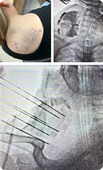
Case Study 1: Pelvic Congestion Syndrome
Chronic pelvic pain and urinary frequency in a woman – definitive diagnosis and treatment with ovarian vein embolization.
Patient profile
Female, 38 years old, mother of two children.
Symptoms and duration
The patient suffered for over two years from chronic pain in the lower abdomen and lower back, which worsened in the afternoon or after prolonged standing. At the same time, she presented with... Frequent urination, a feeling of pressure in the right hip, dyspareunia (discomfort during sexual intercourse) and occasional... urine leakage during sneezing or intense laughter.The symptoms were so burdensome that they limited her social and professional activities.
For a long time, she had received the wrong diagnosis of... Endometriosis and had undergone treatments without success, which had increased her frustration and anxiety.
Diagnosis
After a new approach, a transvaginal Doppler ultrasound was performed, revealing dilated pelvic veins. The final confirmation was made with... Phlebography, which revealed extensive blood reflux.
Therapeutic approach
The minimally invasive treatment with endovascular embolization of the ovarian veins was decided
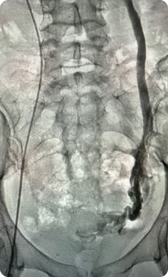
Description of the procedure
The procedure was performed under local anesthesia, through catheterization from the right femoral vein. Under fluoroscopic guidance, the pathological veins were catheterized, and coils were placed along with a sclerosing agent. The entire procedure lasted approximately 40 minutes, without complications.
Course and recovery
The patient was mobilized immediately and discharged on the same day. By the first week, she reported... a clear reduction in pain, and after 6 months of follow-up, the symptoms had completely disappeared. Her quality of life improved dramatically, with a return to daily activities.
What made it unique?
The long-standing incorrect diagnosis and the failure of other treatments highlighted how often Pelvic Congestion Syndrome remains undiagnosed. The case emphasized the importance of a proper diagnostic approach.
Message to the public:
Chronic pelvic pain is not always a gynecological issue. Specialized vascular surgical assessment and modern minimally invasive techniques can offer a definitive solution.